|
Secondary school Biology seldom go into the details, the structure of a cell is an example of this. We were taught that cells consists of the nucleus, mitochondria, chloroplasts, walls and such, but that's pretty much it. We bet that teachers could go deeper into the subject with no risk of confounding their students. We therefore challenge the teachers to add value to their syllabus, starting from the basics.
Cikgus, make us proud and prove us right. |
"Every neuron contains about a million pumps—each one is a slight bump on the cell membrane—and every pump can swap about 200 sodium ions for 130 potassium ions every second." - Anthony Snith, "The Mind", 1986
The cell membrane is a thin flexible layer around the cells of all living things. It is sometimes called the plasma membrane or cytoplasmic membrane.
Its basic job is to separate the inside of cells from the outside. In all cells, the cell membrane separates the cytoplasm inside the cell from its surroundings. In animal cells, that is all there is. Bacteria, fungi and plants have cell walls as well, which support the cell and block the passage of large molecules. This selectively-permeable membrane controls the exchange of materials, receives hormone messages and is very thin. It can be described as a phospholipid bi-layer - meaning that it is made from phospholipid molecules and has two layers.
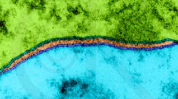
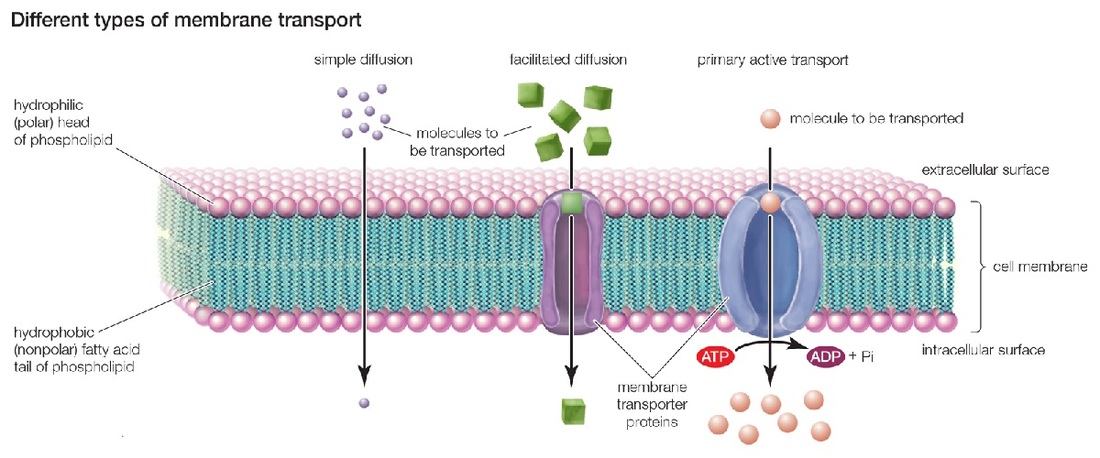
This two layers of phospholipid molecules with phosphate heads on the surfaces and lipid (oil) tails on the inside. The outside heads mix with water, but the tails reject water. The phospholipid bi-layer is so thin it can barely even be seen by an electron microscope - a x100,000 magnification is required, and only shows a double black line around 7 nm wide. Since we cannot properly see the membrane, we have to take what we know about it and create a model - in this case known as a fluid mosaic model.
Its basic job is to separate the inside of cells from the outside. In all cells, the cell membrane separates the cytoplasm inside the cell from its surroundings. In animal cells, that is all there is. Bacteria, fungi and plants have cell walls as well, which support the cell and block the passage of large molecules. This selectively-permeable membrane controls the exchange of materials, receives hormone messages and is very thin. It can be described as a phospholipid bi-layer - meaning that it is made from phospholipid molecules and has two layers.
This two layers of phospholipid molecules with phosphate heads on the surfaces and lipid (oil) tails on the inside. The outside heads mix with water, but the tails reject water. The phospholipid bi-layer is so thin it can barely even be seen by an electron microscope - a x100,000 magnification is required, and only shows a double black line around 7 nm wide. Since we cannot properly see the membrane, we have to take what we know about it and create a model - in this case known as a fluid mosaic model.
Fluid Mosaic Model
As the plasma membrane is so thin that its existence and structure was originally credited to circumstantial evidence until the transmission electron microscope revealed the filmy nature of this lipid bilayer. One form of evidence for the cell membrane involved the observation that oils and hydrophobic solvents passed easily into cells. Knowing this researchers postulated that the membrane must have a lipid core.
The Fluid Mosaic Model refers to the fluidlike qualities of the phospholipid sheets and the dynamic behavior of proteins that seem to float in or on a "sea" of phospholipids. These phospholipids have a polar head group and a nonpolar hydrocarbon chains that resembles a forked tail. The polar region is hydrophilic because of the positively charged variable group attached to the phosphate, whereas the nonpolar fatty acid tails are hydrophobic.
These molecules can move about by diffusion in their own layer with a width of about 7 nm on average. Some of the phospholipids are saturated and some are unsaturated, and this affects the fluidity of the membrane, a heavily unsaturated membrane means a more fluid membrane. This is due to the kink in saturated tails causing the molecules to not sit closely together. The Phospholipid tails point inwards, facing each other, meaning that inside the membrane it is non-polar hydrophobic. The protein molecules within the structure can move around although some are fixed to structures inside the cell and do not move. Also, some of them span the width of the membrane, some are only on the inner layer and some on the outer layer. Many proteins and lipids have short carbohydrate chains attached to them, forming glycoproteins and glycolipids.
Transport
Transport across the phospholipid bi-layer is regulated, and it is an effective barrier - but exchange is necessary. Methods of exchange are in terms of passive transport, active transport and bulk transport.
Osmosis is the movement of water through a plasma membrane from a region of low solute concentration to a region of high concentration. Osmosis is passive transport through diffusion, meaning it does not require energy to be applied. What causes osmotic pressure is different concentrations of solutes on the two sides of the membrane.
Active transport is defined as the energy-consuming transport of molecules or ions across a membrane against a concentration gradient, made possible by transferring energy from respiration. The energy is supplied by ATP, and is used to make the transport protein change its shape, transferring the molecules or ions across the membrane in the process. Something to note is that cells that do a lot of active transport are likely to have many mitochondrion to provide the energy for it.
It is particularly important in reabsorption in the kidneys where certain useful molecules must be reabsorbed into the blood after filtration. In plants it is used to load sugar from photosynthesising cells into the phloem tissue for transport.
Bulk transport can be defined as the movement of large quantities of materials into or out of cells, endocytosis and exocytosis, respectively.
Exocytosis is the process by which materials are removed from cells - for example the secretion of digestive enzymes, where vesicles from the Golgi apparatus carry the enzymes to the cell surface, bind to it, and release their contents.
Endocytosis is the reverse of exocytosis and involves the engulfing of the material by the cell to form a small sac inside the cell. The most common form is phagocytosis, performed by phagocytes - an example of which would be white blood cells engulfing bacteria. The second form of endocytosis is pinocytosis, the bulk uptake of liquid, and the human egg cell take sup nutrients from cells that surround it by this method.
Example: Mammalian Lungs
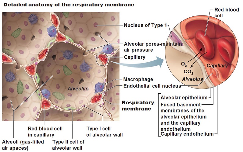
All mammals need a supply of oxygen to use in respiration - and in mammals the cells that require the oxygen are too far for diffusion to be effective and so they have a specialised gaseous exchange surface, where oxygen can diffuse into the body and carbon dioxide can diffuse out. In humans, this exchange surface is the alveoli in the lungs, and as you can see in the diagram below, each alveolus is tiny but collectively they have a huge surface area. In an average human lung, there are 480 million alveoli, totaling around 70m2 in an adult. This increases the net rate of diffusion. Alveoli also have extremely thin walls, no more than 0.5 μm thick, directly next to blood capillaries also with very thin cell membranes. This thinness allows diffusion to be extremely speedy.
When a mammal breathes in, the concentration of oxygen is higher in the alveolus than in the red blood cells. Therefore, oxygen leaves the alveolus and enters the red blood cells. When a mammal breathes out, the opposite happens. The concentration of carbon dioxide is lower in the alveolus than in the red blood cells, so carbon dioxide leaves the red blood cells, enters the alveolus, and is exhaled.
Since diffusion only works down a concentration gradient, and the steeper said concentration gradient, the faster diffusion, the concentration gradient between the alveoli and the blood must be kept steep constantly to ensure speedy diffusion. Carbon dioxide is rapidly breathed out, meaning that carbon dioxide transported to the alveoli will be quickly transported into them and breathed out, and deoxygenated blood is constantly kept coming to the alveoli, meaning that oxygen diffuses into it quickly.
Ponder this
Why are cell membranes lipid (fat) based? What are the advantages and disadvantages of a carbohydrate-, or protein-based membrane?
As osmosis can both be advantageous and disadvantageous (hyper/hypo hydration), would it not be better if cell membranes are water-proof, and depended on active transport for osmotic balance?
Discuss
How do variations in the fluid mosaic model structure account for the functional differences among membranes? How does the structural organization of membranes provide for transport and recognition? How are the structures of the various subcellar organelles related to their functions? Do these factors limit cell size? Why or why not?
Further reading
Cell membrane, at Wikipedia
Components and structure in the Fluid Mosaic Model, at the University of California - Davis' Biowiki
History of the cell membrane theory, a look at the formulation and development of membrane theory throughout history